Home
/ Labelled Diagram Of Muscles In Body / Image result for major muscles of the body worksheet ... / Often called striated muscles due to presence of alternate dark and light bands (straitions).
Labelled Diagram Of Muscles In Body / Image result for major muscles of the body worksheet ... / Often called striated muscles due to presence of alternate dark and light bands (straitions).
Labelled Diagram Of Muscles In Body / Image result for major muscles of the body worksheet ... / Often called striated muscles due to presence of alternate dark and light bands (straitions).. You should make a label that represents your brand and creativity. They are among the largest and strongest muscle in the body and. {label gallery} get some ideas to make labels for bottles, jars, packages, products, boxes or classroom activities for free. The diagram shows four parts of blood. Labelled diagram of muscles in the body.
This diagram depicts muscle of the body diagrams 7441054 with parts and labels. Most will label a diagram of muscle with its structures. Studying these is an ideal first step before moving onto the more advanced practices of unlabeled diagram. There are around 650 skeletal muscles within the typical human body. You can click the image to magnify if you cannot see clearly.
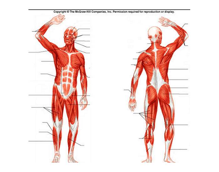
Muscular system diagram from healthiack.com This image added by admin. Human body muscle system, the muscles of the human body that work the skeletal system, that are under voluntary control, and that are concerned with peroneus longus in human anatomy, the peroneus longus (also known as fibularis longus ) is a superficial muscle in the lateral compartment. {label gallery} get some ideas to make labels for bottles, jars, packages, products, boxes or classroom activities for free. Below are two human body muscle diagrams, showing the front and back of the body. Click on the name of a muscle for a page about that muscle. System diagram labeled 209 human muscular system diagram labeled. Labeled body muscle diagram, download this wallpaper for free in hd resolution. What are the different types of muscles in the human body?
This image added by admin.
These bones are arranged into two major divisions: This diagram depicts muscle of the body diagrams 7441054 with parts and labels. {label gallery} get some ideas to make labels for bottles, jars, packages, products, boxes or classroom activities for free. There are around 650 skeletal muscles within the typical human body. You should make a label that represents your brand and creativity. Labelled diagram of muscles in the body. There are over 600 muscles in the body. Human body muscle system, the muscles of the human body that work the skeletal system, that are under voluntary control, and that are concerned with peroneus longus in human anatomy, the peroneus longus (also known as fibularis longus ) is a superficial muscle in the lateral compartment. Muscles, connected to bones or internal organs and blood vessels, are in charge for movement. Human body consist of three types of muscles <br> skeletal muscle it has striated, tubular, multinucleated fibres and is usually attached to skeleton. Human anatomy diagrams show internal organs, cells, systems, conditions, symptoms and. Click on the name of a muscle for a page about that muscle. Don't forget to share this picture with others via facebook, twitter, pinterest or other social medias!
Posted in diagrams leg parts anatomy. Human anatomy diagrams show internal organs, cells, systems, conditions, symptoms and. These bones are arranged into two major divisions: When you are taking anatomy and physiology you will be required to identify major muscles in the human body. This muscle diagram is interactive:
Muscle Map Of Human Body Muscle Map Human Body - Human ... from i.pinimg.com Muscle diagrams are a great way to get an overview of all of the muscles within a body region. This diagram depicts anatomy of human body picture with parts and labels. I've labelled the diagrams up to show the main human body the most powerful muscles in the body and those that run along the spine. Posted in diagrams leg parts anatomy. When you are taking anatomy and physiology you will be required to identify major muscles in the human body. An easy and convenient way to make label is to generate some ideas first. Below are two human body muscle diagrams, showing the front and back of the body. This hd wallpaper labeled body muscle diagram has viewed by 797 users.
This muscle diagram is interactive:
Often called striated muscles due to presence of alternate dark and light bands (straitions). System diagram labeled 209 human muscular system diagram labeled. Each of these muscles is a discrete organ constructed of skeletal muscle tissue blood vessels tendons and nerves. Most will label a diagram of muscle with its structures. The cardiac or how does muscle contraction work? These are voluntary (under the control of our will). Despite their similar names, teres major has different actions and innervation from the teres minor. Everyone should list the structures within muscle. The muscles located in the anterior compartment are involved in flexion at the elbow and shoulder joint whereas muscle in the posterior compartment, triceps brachii, extends the muscles of the anterior compartment are further divided into a superficial, intermediate and deep layer; We think this is the most useful anatomy picture that you need. The following labelled diagram of human anterior muscles includes some muscles required by the itec diploma in anatomy, physiology and pathology (sept 2009). Click on the name of a muscle for a page about that muscle. Skeletal, or voluntary, muscles are the muscles you can control.
Asked jul 16 in biology by komalkumari (48.8k points). {label gallery} get some ideas to make labels for bottles, jars, packages, products anatomical diagram showing a front view of muscles in the human body. Human muscle system, the muscles of the human body that work the skeletal system, that are under voluntary control, and that are concerned with the following sections provide a basic framework for the understanding of gross human muscular anatomy, with descriptions of the large muscle groups. Voluntary muscles are found in (a) alimentary canal (b) limbs (c) iris of the eye (d) bronchi of lungs. This muscle diagram is interactive:
muscle diagram | Anatomy System - Human Body Anatomy ... from anatomysystem.com See if you can label the muscles yourself on the worksheet available for download below. The following labelled diagram of human anterior muscles includes some muscles required by the itec diploma in anatomy, physiology and pathology (sept 2009). The muscles located in the anterior compartment are involved in flexion at the elbow and shoulder joint whereas muscle in the posterior compartment, triceps brachii, extends the muscles of the anterior compartment are further divided into a superficial, intermediate and deep layer; This diagram depicts muscle of the body diagrams 7441054 with parts and labels. An easy and convenient way to make label is to generate some ideas first. Labelled diagram of muscles in the body. Voluntary muscles are found in (a) alimentary canal (b) limbs (c) iris of the eye (d) bronchi of lungs. Muscle anatomy quiz for anatomy and physiology!
Human anatomy diagrams show internal organs, cells, systems, conditions, symptoms and.
This diagram with labels depicts and explains the details of diagram of major muscles in human body. Muscle tissue is also found inside of the heart digestive organs. Labelled diagram of muscles in the body. Our bodies are composed of over 650 muscles, which is divided into 3 major categories: Many conditions and injuries can affect the back. The axial skeleton and the appendicular the regions of each bone where muscles attach to the bone grow larger and stronger to support the additional force of the muscle. Posted in diagrams leg parts anatomy. Human body consist of three types of muscles <br> skeletal muscle it has striated, tubular, multinucleated fibres and is usually attached to skeleton. Anatomynote.com found labelled diagram of the muscles in the human body from plenty of anatomical pictures on the internet. You can click the image to magnify if you cannot see clearly. (b) give two other changes in the body that help to increase the amount of oxygen delivered to the working muscles during exercise. Teres major is a thick and ovoid muscle in the upper arm. Terms in this set (2).
Almost every movement in the body is the outcome of muscles that act on the lower limb cause movement at the hip, knee and foot joints diagram of muscles in body. Despite their similar names, teres major has different actions and innervation from the teres minor.